IL-31 regulates DC-mediated T cell responses in the course of atopic dermatitis
Based on previous work from our laboratory, showing that DCs express the IL-31R complex under certain conditions and release a number of pro-inflammatory mediators upon stimulation with IL-31, we assume that IL-31-stimulated DCs contribute to the complex picture of T cell responses in AD. We hypothesize that, in the course of AD, Th0 cells, a type of CD4+ T cell expressing Th1- as well as Th2-cytokines, may serve as a source of IFN-gamma, which we could pinpoint as the activator of IL-31R expression in DCs. Thus, DCs expressing functional IL-31R are responsive to IL-31 stimulation and activate downstream signaling pathways that result in the secretion of pro-inflammatory mediators such as IL-6. These soluble factors (and maybe also the expression of co-stimulatory molecules) can promote the activation of specific CD4+ T cell populations which subsequently release increasing amounts of IFN-gamma, IL-17A and possibly IL-10. Since it has recently been reported that an increase in IFN-gamma- and IL-17-producing cells correlates with the severity of AD), we assume that IL-31, a cytokine that was also shown to be associated with disease severity, might be a key factor in inducing the switch in the cytokine milieu as observed in the progression of AD. 
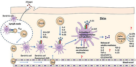
Model of IL-31-induced DC-mediated T cell activation in AD
Upon allergen contact, dendritic cells migrate to the lymph nodes where they present allergen to naive CD4+ T cells. In the presence of IL-4, T cells differentiate toward a Th2 phenotype. IFN-gamma, released by Th1/0 CD4+ T cells induces the expression of IL-31R in DCs. Th2 cells, which are attracted to the site of inflammation (skin), release IL-4, IL-5, IL-13 and IL-31. The latter binds to the IL-31R on DCs and activates specific signaling cascades resulting in the release of pro-inflammatory mediators and the expression of co-stimulatory molecules. This in turn induces the activation of particular Th cell subsets. The subsequent release of cytokines contributes to the progression of AD.
 This project is funded by the Austrian „Fond zur Förderung der Wissenschaftlichen Forschung“, Grant P23933.


