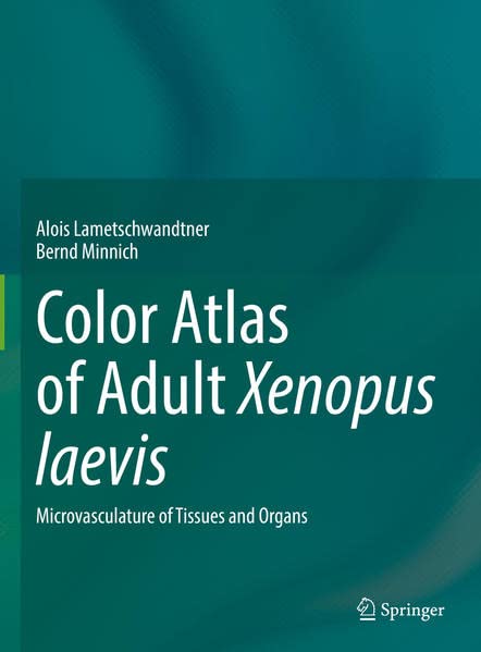
Color Atlas of Adult Xenopus laevis. Microvasculature of Tissues and Organs
Color Atlas of Adult Xenopus laevis
Microvasculature of Tissues and Organs
- Comprises more than 300 colored figures
- Offers a detailed account on the circulatory system of Xenopus laevis
- Sets a particular emphasis on the tissue- and organ-specifc microvascular anatomy
This atlas offers stunning color electron scanning micrographs and exceptional light microscopy pictures of capillaries, vessels and diverse histomorphological tissues and organs of Xenopus laevis. The model organisms Xenopus laevis serves to study basic biological questions related to growth, differentiation, maturation, and regression of cells, tissues and organs. Xenopus and human genomes have long stretches of gene collinearity, and 79% of identifed human disease genes have a verifed ortholog in Xenopus. Thus, this atlas will be a powerful tool for anatomists, morphologists, histologists and physiologists interested in normal and pathologically altered organs and tissue; and to all researchers, who wish to learn more about the microvascular anatomy of this vertebrate model organism.
Color Atlas of Adult Xenopus laevis | SpringerLink


