Single-celled plankton reveal a new source of protein-based biomaterials
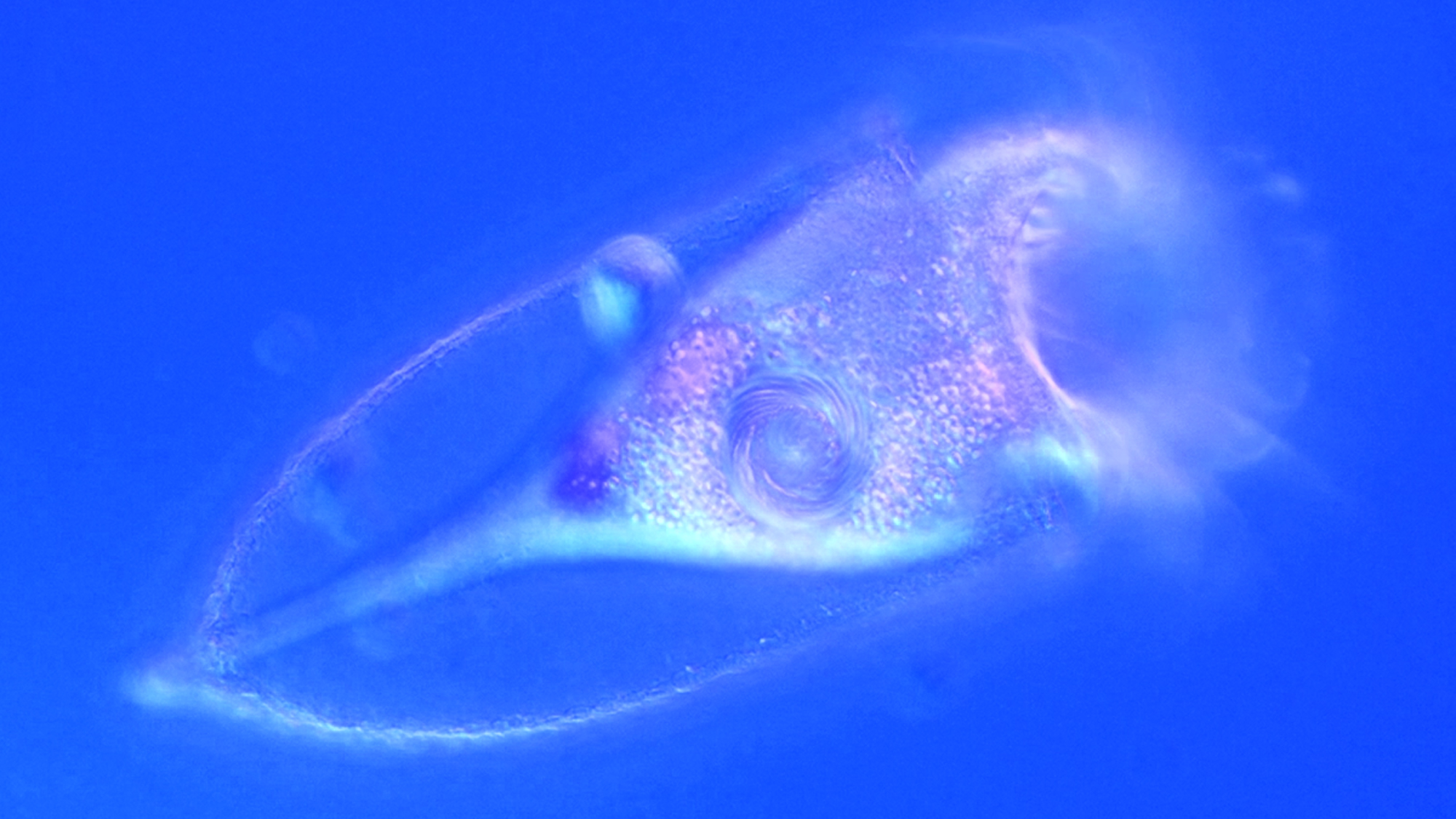
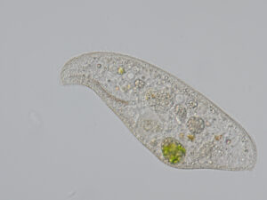
Imagine a single minute cell building a transparent vase-shaped shell around itself. It has no hands, no spinning organ, and no nervous system. Yet, within minutes after cell division, a tintinnid ciliate can release material into the surrounding water, which forms autonomously an intricate protective shell.
For more than 200 years, biologists have admired these shells under the microscope. Tintinnids are planktonic ciliates, single-celled eukaryotes that live in marine and freshwater, and their shells can look like tiny tubes, cups, or works of glass art. They are beautiful, but they are also remarkably tough. Tintinnid shells can withstand exterme heat and harsh chemical treatments that would destroy many other biological structures.
Living tintinnid in its transparent shell during cell division. Credit: Maximilian Ganser
That raised a simple question with a surprisingly difficult answer: What are these shells made of?
In our recent study, we found that tintinnid shells are composed of self-assembling structural proteins. We named this newly discovered protein family Tintinnidorin because, it appears to be unique to tintinnid ciliates.
This finding matters because many of the best-known protein-based biomaterials come from animals. Spider silk, for example, is famous for combining strength, elasticity, and lightness. Silkworm silk has been used by humans for thousands of years. These materials inspire research in textiles, medicine, and engineering, but the organisms that produce them are complex animals with specialized organs and highly controlled production systems.
Tintinnids offer a very different model. They are single cells. They simply secrete their shell-forming material into water, where it organizes into a stable structure. That makes them especially interesting for understanding how proteins can store information not only for biological function, but also for material formation.
To solve the mystery, we combined methods from several fields. We analyzed the proteins present in tintinnid shells and compared them with genetic information from tintinnid cells. In other words, we looked for a match between the material outside the cell and the molecular blueprints inside the cell. This combination of shell proteomics, single-cell transcriptomics, and bioinformatics allowed us to identify the protein sequences that form the shell.
The result was unexpected. Tintinnidorin proteins are unlike ordinary globular proteins that fold into one compact structure. Instead, they appear to combine folded regions with flexible segments. This architecture may help explain how the proteins can be stored inside the cell, secreted into the environment, and then assemble into a resistant shell. Their composition also helps explain properties such as stability, wet adhesion in salty water, and the ability to absorb ultraviolet light.
For biomaterials research, this is exciting because it expands the search space. Nature has already evolved many protein materials with properties that engineers would like to reproduce: strength, flexibility, chemical resistance, adhesion under wet conditions, and sustainable production. Until now, most attention has focused on animals. Our study shows that eukaryotic single-celled organisms can also produce sophisticated structural biomaterials.
Tintinnids are also diverse. About 1,000 species are known, differing in the shapes, textures and architectures of their shells. This diversity suggests that Tintinnidorin proteins may also be diverse. We have likely seen only the beginning of what this group of proteins can do.
There is still much to learn. We do not yet know all the rules that connect Tintinnidorin sequences to the final shell structures. Current protein structure prediction tools are optimized mainly for globular proteins, so unusual structural proteins like Tintinnidorins challenge our existing methods. However, that challenge is also an opportunity. By studying tintinnids, we can learn how single cells build stable materials from proteins and perhaps use those principles to design new sustainable materials in the future.
For us, the most fascinating part is that this discovery came from organisms that are easy to overlook. Tintinnids are microscopic plankton, but they have solved a materials problem that humans are still trying to understand: how to build complex, resilient structures from simple biological components in water.
Their shells remind us that innovation in nature is not limited to large animals, plants or familiar model organisms. Sometimes, a major clue to future biomaterials science is drifting invisibly in a drop of seawater.
- Maximilian H. Ganser et al. Self-assembling proteins compose the chemically resistant shell biomaterial of planktonic tintinnid ciliates, Nature Communications (2026). DOI: 10.1038/s41467-026-74402-4
Pupil internship financially supported by FFG
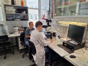 Thomas Weißenbacher from the AHS Hallein spent four weeks in the research group “Protistology”. He gained an overview of the field of biology, with a focus on unicellular organisms. Further, Thomas became familiar with various protist groups as well as with the different investigation methods and their results and learned to apply some of them. He was introduced into the key questions of the current research project on shells (loricae) of tintinnid ciliates from the marine plankton. Thomas got insights into the numerous complex steps involved in preparing samples for transmission electron microscopy, as well as into the fundamentals of electron microscopy. This knowledge he applied in the analyses of transmission electron microscopic images. Additionally, Thomas learned how to maintain a lab notebook. The sampling of freshwater plankton (qualitatively and quantitatively) plus establishing ciliate cultures were other topics of his internship. Thomas Weißenbacher became capable of operating a stereomicroscope and a microscope and identified the collected organisms, using different identification keys. Eventually, he established a species list illustrated with his own photomicrographs. After receiving a training in the general and specific laboratory regulations, Thomas learned to demonstrate water currents generated by protists, to detect pH changes during the digestion of food particles in unicellular organisms, and to stain the cell nuclei and ciliature. The instructional units were based on PowerPoint presentations and custom educational films which provided the theoretical foundation for the practical work and always preceded the different work phases.
1st place for Laura Böll at international protistology conference
Laura Böll, Bachelor student of the working group of Assoc. Prof. Dr. Sabine Agatha / Protistology at the Department of Environment & Biodiversity, presented her poster at the 43rd conference of the German Society for Protozoology. Among the fascinated participants from Germany, Austria, Switzerland, Great Britain, the Netherlands, France and Poland, her work was awarded the coveted 1st place.
As part of a unique FWF project on tintinnids, the shell-building ciliates of marine plankton, Laura Böll intensively investigated the production of the building material for the vase-shaped shells. For the first time, she was able to determine the time of material production in the cell cycle and quantify the amount of substance available to the anterior of the two dividers for construction. The result: the intracellular material swells massively after secretion and the finished housing wall is therefore significantly more voluminous.
Further fascinating results were presented by Dr. Maximilian Ganser, also from Sabine Agatha’s working group / Protistology. The aim of his investigations was to identify tintin-specific genes from the transcriptomes of individual cells, which are necessary for the construction of the shell, among other things, and are therefore absent in the shell-less closest relatives.
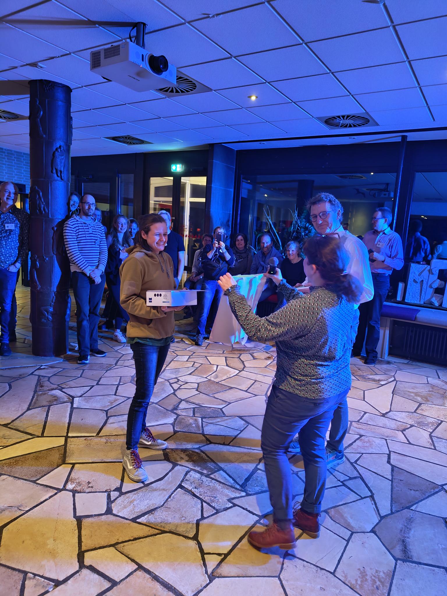
Laura Böll at the award ceremony of the German Society for Protozoology in Haltern am See, Germany. Photo: Maximilian Ganser
“And unfortunately over again – the marine biology excursion to Rovinj, Croatia”
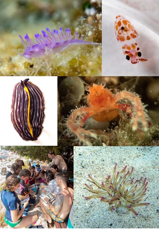
From July 16 to 22, 2023, 18 Bachelor students went on an excursion to Rovinj, Croatia, as part of the Marine Biology module. They were accompanied by Assoc. Prof. Sabine Agatha, Anna Oberlercher and Kilian Niedermeyer. In addition to the familiar variety of fish, crustaceans, echinoderms, sponges, algae etc., new highlights were found this year: the nudibranch Flabellina elegans, young cuttlefish (Sepia officinalis; approx. 1 cm long), the flatworm Prostheceraeus giesbrechtii, the crab Munida tenuimana and the anemone Condylactis aurantiaca. Getting to know organisms from different habitats (sediment, hard bottom, plankton) and understanding the adaptations of the organisms to these habitats were the focus of the snorkeling excursions to various spots on the offshore islands and along the coast. The field work was supplemented by sampling with the institute’s ship and units in the course room.
Congratulations to Dr. Maximilian Ganser
Congratulations to Dr. Maximilian Ganser on the successful defence of his PhD Thesis “An Integrative Approach to Marine Planktonic Ciliates (Alveolata, Ciliophora, Oligotrichea): Novel Aspects of their Diversity, Evolution, and Geographic Distribution”. Committee members were Assoc. Prof. Dr. Micah Dunthorn, Natural History Museum of the University of Oslo, and Assoc. Prof. Dr. Peter Vďačný, Department of Zoology at the Comenius University in Bratislava.
His thesis contained five peer-reviewed papers:
- Ganser, M.H., Agatha, S., 2019. Redescription of Antetintinnidium mucicola (Claparède and Lachmann, 1858) nov. gen., nov. comb. (Alveolata, Ciliophora, Tintinnina). J. Eukaryot. Microbiol. 66, 802-820.
- Ganser, M.H., Forster, D., Liu, W., Lin, X., Stoeck, T., Agatha, S., 2021. Genetic diversity in marine planktonic ciliates (Alveolata, Ciliophora) suggests distinct geographical patterns – data from Chinese and European coastal waters. Front. Mar. Sci. 8, 643822.
- Ganser, M.H., Santoferrara, L.F., Agatha, S., 2022. Molecular signature characters complement taxonomic diagnoses: A bioinformatic approach exemplified by ciliated protists (Ciliophora, Oligotrichea). Mol. Phylogenet. Evol. 170, 107433.
- Agatha, S., Ganser, M.H., Santoferrara, L.F., 2021. The importance of type species and their correct identification: A key example from tintinnid ciliates (Alveolata, Ciliophora, Spirotricha). J. Eukaryot. Microbiol. 68, e12865.
- Hütter, T., Ganser, M.H., Kocher, M., Halkic, M., Agatha, S., Augsten, N., 2020. DeSignate: detecting signature characters in gene sequence alignments for taxon diagnoses. BMC Bioinformatics 21, 151.
Protists from the 3D-printer, 1st June 2022
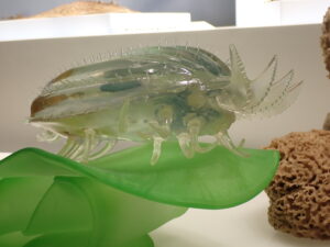
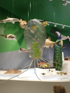
The recently opened exhibition of the Haus der Natur displays two new models of protists at 5,000 to 6,000× magnification ( https://www.hausdernatur.at/de/). The models of a ciliate (Ciliophora) of the genus Euplotes and of a testate amoeba (Cercozoa) of the genus Euglypha were created in close collaboration with the research group Protistology of Assoc. Prof. Dr. Sabine Agatha. The 3D models have prominent position in the showcase “Biodiversity” and demonstrate impressively the complexity of unicellular organisms. Actually, these single cells are consummate organisms that perform locomotion, feed, digest, defecate, proliferate, and even have sex. The numerous genders of ciliates necessitate a particularly elaborate mate selection by means of pheromones.
Lange Nacht der Forschung (Long Night of Science), 20th May 2022
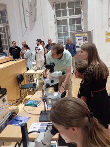
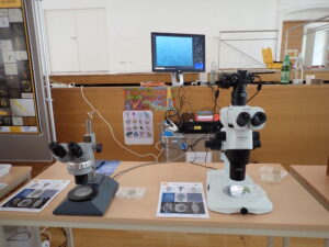
The “Long Night of Science” at the Faculty of Catholic Theology and in the assembly hall of the University of Salzburg was extremely successful. The research group Protistology presented many visitors a huge variety of microscopic organisms living in freshwater, mainly unicellular organisms (protists) with bizarre lifestyles (Courses).
City Nature Challenge, April 2022
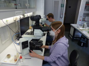
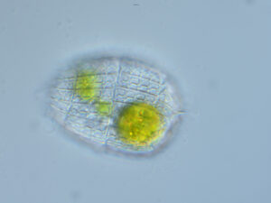
The research group Protistology and several interested students microscopically studied and identified numerous protists and mesozooplankton organisms from puddles, ponds, and lake in Salzburg and added them to the records of the City Nature Challenge. Finally, Salzburg compiled 12.653 observations including microscopic organisms. Thereby, Salzburg became second best in Austria and Europe and was among the top 5% worldwide.

Cover picture of scientific journal, January 2022
The 3D reconstruction of the somatic infraciliature by Michael Gruber, former PhD student in the research group Protistology, is cover picture of the international peer-reviewed Journal of Eukaryotic Microbiology published by Wiley.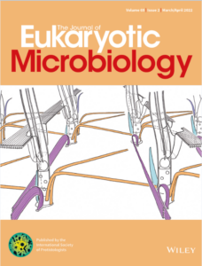
Sabine Agatha among the World’s top 2 per cent of Scientists
The team of John P. A. Ioannidis from the Stanford University recently published a ranking of the world’s top 2% of scientists (Ioannidis JPA, Boyack KW, Baas J (2020) Updated science-wide author databases of standardized citation indicators. PLoS Biol 18(10): e3000918. https://doi.org/10.1371/journal.pbio.3000918). They assessed the citation impact during the calendar year 2019 (Table-S7-singleyr-2019) and listed S. Agatha among the top 2%.
Publication of the Month – May 2020 for Maximilian Ganser and Thomas Hütter
“DeSignate: detecting signature characters in gene sequence alignments for taxon diagnoses”Maximilian H Ganser, Thomas Hütter, Manuel Kocher, Merima Halkic, Sabine Agatha & Nikolaus Augsten
Abstract: Species of prokaryotes and eukaryotes including plants, animals, fungi, and unicellular protists are described and named by taxonomists. These descriptions include species diagnoses which state all characters distinguishing a species from its closest known relatives. Diagnostic characters mainly comprise morphological differences while molecular differences are rarely included because efficient solutions are lacking. We therefore developed the software tool DeSignate in an interdisciplinary project between the Protistology Research Group (AG Agatha, Biosciences) and the Database Research Group (AG Augsten, Computer Sciences). DeSignate enables the detection and classification of diagnostic molecular characters in a user-friendly web interface which can subsequently be included in diagnoses. For the program see, https://designate.dbresearch.uni-salzburg.at/.

Introduction into Electron Microscopy for Pupils from Grammar School Zaunergasse
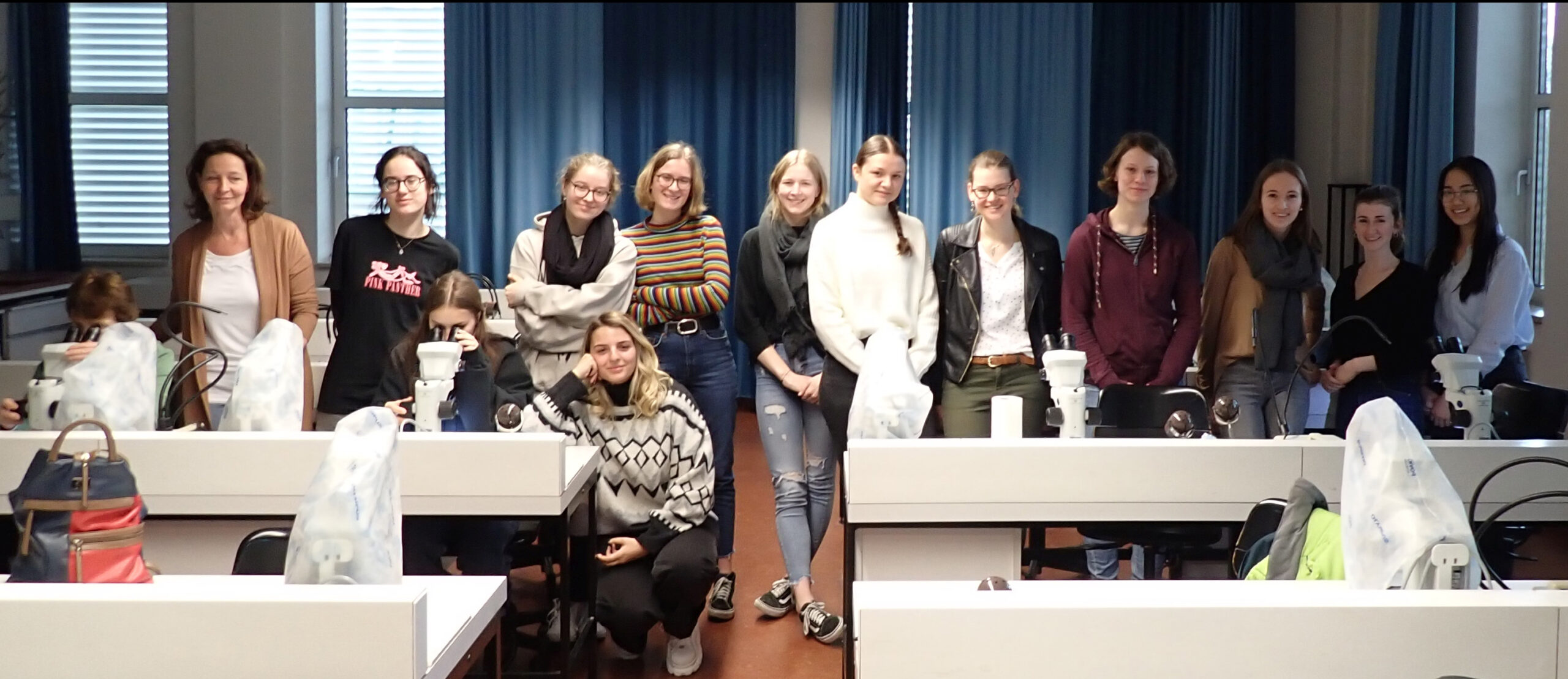
Two groups of pupils from the grammar school in the Zaunergasse in Salzburg visited together with their teacher Leopoldine Sams the Department of Biosciences on February 18th and 19th. Prof. Ulrike Berninger gave a short overview of the biological studies at the University of Salzburg. Then, Assoc.-Prof. Dr. Sabine Agatha and Mag. Birgit Weissenbacher from the working group ‘Protistology’ gave an introduction into scanning and transmission electron microscopy. The functioning of the electron microscopes and the time-consuming preparation of the samples, using sophisticated techniques and facilities, was demonstrated in own videos. A tour through the department with a detailed demonstration of the electron microscopical facilities completed the visit. All participants, the teacher, and the staff of the Department of Biosciences had a lot of fun.
New Videos
Videos on preparations for scanning and transmission electron microscopy of protists as well as on live observation of the model ciliate Paramecium sp. were produced by the AG Agatha; they are available under “Lehre“.
Poster price for Michael Gruber at Meeting of NOBIS Austria
At the 13th annual meeting (November 28th & 29th 2019) of the Network of Biological Systematics (NOBIS) Austria in Salzburg, the poster of Michael Gruber with the title “Tintinnid’s first top model: ultrastructural insights into the oral ciliature of Schmidingerella meunieri (Alveolata, Ciliophora)” was rewarded.

Awardees: Susanne Affenzeller (best talk of a PhD student), Michael Gruber (AG Agatha; best poster), and Silvester Bartsch (best talk of a Master student).
With three talks and four posters at the European Congress of Protistology in Rome
The European Congress of Protistology takes place every four years and participants come not only from European countries, but also from North and South America, China, Japan, Korea, and India to name only the most dominant ones. This year, the congress took place in Rome, Italy, from July 28th to August 2nd. Sabine Agatha, Maximilian Ganser, and Michael Gruber presented their recent results in three talks (incl. one plenary talk) and three posters covering several mainly ultrastructural aspects of tintinnid ciliates, viz., the oral and somatic ciliature, the resting cyst, the extrusomes (capsules), and the lorica walls. Additionally, the findings on the diversity of tintinnids from the Northeast Pacific including supposedly new species was shown.
Our article is one of the top downloaded!
Our article “Redescription of Tintinnopsis everta Kofoid and Campbell 1929 (Alveolata, Ciliophora, Tintinnina) Based on Taxonomic and Genetic Analyses-Discovery of a New Complex Ciliary Pattern”, published in the Journal of Eukaryotic Microbiology (65: 484–504; doi: 10.1111/jeu.12496), is one of the journal’s top downloaded recent papers! This means: Amongst articles published between January 2017 and December 2018, our paper received some of the most downloads in the 12 months following online publication. Thus, our work generated immediate impact and visibility, contributing significantly to the advancement of your field.
“Microscopic Life in Soil” Unterrichtseinheiten für die 6. Klasse AHS Oberstufe konzipiert von Frau Mag. rer. nat. Susanne Riedl
Protozoen beeinflussen die Bodenfruchtbarkeit und somit die Nahrungsverfügbarkeit für den Menschen, indirekt über den Einfluss auf die Bakterienpopulation, die Hauptzersetzer der Streu, und direkt durch die Abgaben von Stickstoffverbindungen. Im Gegensatz zu Schulbüchern, welche sich üblicherweise auf Metazoen fokussieren und dabei die Protozoen außer Acht lassen, führt die vorliegende Diplomarbeit Unterrichtseinheiten ein, welche sich auf die spezifischen Anpassungen an den ephemeren Charakter des Bodens sowohl der Protozoen als auch der Metazoen konzentrieren und einen vereinfachten Einblick in das Bodennahrungsnetz liefern. Trotz ihrer mikroskopischen Größe sind Protozoen, insbesondere Ciliaten, einfach zu handhaben und liefern genügend morphologische Kennzeichen zur Unterscheidung der Hauptgruppen. Dementsprechend ist es für die Schülerinnen und Schüler möglich, ihre eigenen Hypothesen in den praktischen Einheiten bezüglich der Sukzession, des Nahrungsnetzes, dem Einfluss von Streu und der Wasserverfügbarkeit auf die Abundanz der Protozoen im Boden zu prüfen. Durch die Abdeckung dieser Themen fügen sich die Einheiten nicht nur perfekt in den Lehrplan der österreichischen höheren Schulen, sondern bieten auch die Möglichkeit eines aktiven und anschaulichen Erwerbens von Wissen und Kompetenzen. Mit Ausnahme der Mikroskope sind die Utensilien für die Einheiten kostengünstig und einfach zu beschaffen; die überwiegend angewandte Methode ist die „non-flooded petri dish“ Technik. © Susanne Riedl